Prodding leukemia cells with nanoprobes could provide cancer clues
02 Jun 2016 by alisonhadley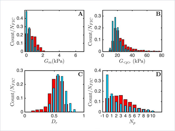 Figure 2 (graphs A, B, C, D). Deformation data: In the study, the team analysed 1672 force indentation curves collected
from 60 healthy cells (represented by the blue, narrow bars) and compared these results with 1302 force indentation curves collected from 51 cancerous cells (represented by the
red, wide bars). (Françoise Argoul et al 2016 Phys. Biol. 13 03LT01).
Figure 2 (graphs A, B, C, D). Deformation data: In the study, the team analysed 1672 force indentation curves collected
from 60 healthy cells (represented by the blue, narrow bars) and compared these results with 1302 force indentation curves collected from 51 cancerous cells (represented by the
red, wide bars). (Françoise Argoul et al 2016 Phys. Biol. 13 03LT01).
Miniature mechanical testers have the potential to chart cell degradation in the body
Giving blood cells a gentle squeeze can reveal a great deal about their health. To find out more, researchers in France have used a tiny force probe to compare the mechanical responses of healthy and cancerous hematopoietic cells (biological structures that help to renew blood in the body).
Publishing their results in the journal Physical Biology, the scientists report that cells harvested from the bone marrow of five leukemia patients appeared much stiffer than comparable samples taken from five healthy volunteers. Also, thanks to the sensitivity of the measurement apparatus – an atomic force microscope – the group was able to identify areas of localized brittle failure accompanying the stiffening of the cancerous cells.
“What makes this work so exciting to us is not simply seeing a difference between the stiffness of healthy and cancerous cells, but observing that the cancerous cells also lost their dynamic ductility and behaved as more breakable objects,” commented Françoise Argoul, who led the study and is a member of the French National Centre for Research (CNRS).
Future applications
The mechanical signatures obtained by squeezing or deforming cells could potentially assist physicians in determining the presence of cancers such as leukemia in patients. Significantly, the mechanical data might also provide clues as to how long the cells have been affected by the disease.
“We would like to construct a hematopoietic cancer cell chart where the loss of cell mechanical functions could be graded, depending on the leukemia and its stage of evolution,” explained Argoul.
Thinking about how the technique might be applied in a hospital setting, she added that biopsy needles could, in principle, be adapted to allow local sensing of internal soft tissue structures.
Validating the approach
Before progressing to testing cells inside the body and preparing for clinical trials, the researchers must first build up sufficient information from their measurements on isolated cells under a range of conditions in the lab.
The team was able to communicate its results rapidly to the scientific community by selecting the Letters publication option offered by the IOP Publishing journal Physical Biology.