Researchers move a step closer to first full-body PET scanner
28 Feb 2017 by Simon Davies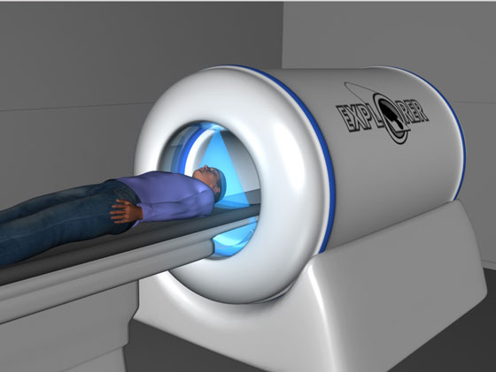 An artist's rendering of the EXPLORER full-body PET scanner. Photo: EXPLORER Project.
Researchers from the University of California, Davis (UC Davis) have revealed the first results from the EXPLORER project – which aims to build world’s first full total-body positron emission tomography (PET) scanner.
Positron emission tomography (PET) is a molecular imaging technique widely used in clinical diagnostics and clinical research to observe metabolic processes and molecular pathways in the body. Current systems, however, are limited in the area of the body they can scan at any one time, which limits their use and does not make best use of the radiation doses involved. The EXPLORER project aims to solve these problems.
Simon Cherry, Professor of Biomedical Engineering at UC Davis and project co-leader said: “The vision of the EXPLORER project is to solve two fundamental limitations of PET as it is currently practiced. The first is to allow us to see the entire body all at once. The second huge advantage is that we’re collecting almost all of the available signal, which means we can acquire higher quality images, or collect the same images as today’s scanners much more quickly or at a much lower radiation dose.
“The EXPLORER scanner will allow all tissues and organs to be imaged simultaneously, with 40 times more sensitivity than the current generation of clinical PET/CT scanners.”
The team’s first paper, published in the journal Physics in Medicine and Biology, reports on development of an efficient image reconstruction method with quantitative correction for the EXPLORER.
Running at full capacity, EXPLORER would be capable of generating around 40 terabytes of data a day, posing a huge challenge for data handling and image reconstruction. The team therefore aimed to develop a quantitative image reconstruction method for the EXPLORER, and compare its performance with current whole-body scanners.
The team, led by Jinyi Qi, a Professor of Biomedical Engineering at UC Davis, used computer simulations to evaluate the image quality, having mimicked a 20-minute whole-body PET scan, using an anthropomorphic phantom with an injection of 25 megabecquerel dose of fluorodeoxyglucose – a standard radiotracer for PET imaging.
They then compared the performance of the EXPLORER with a current clinical scanner. The simulation study showed the EXPLORER could reach superior performance for total-body PET imaging at very low radiation doses, with a six-fold increase in the signal-to-noise ratio compared with the current scanner.
Ramsey Badawi, Professor of Radiology and co-leader of the project said: “In essence, what we aimed to do was test if it possible to efficiently process all the data generated from the half a million detectors needed for a total-body scanner.”
“Our simulations show EXPLORER will enable PET imaging studies of the whole body to be performed using 1/40th the radiation dose, or in 1/40th of the time (potentially in a single breath-hold), or with 40-fold better signal.”
“At 1/40th of the radiation dose, the radiation exposure is equivalent to what you would receive on a round-trip transatlantic flight. All of this means it’s going to have some profound implications for how we use PET scanning in medicine and medical science.”
The EXPLORER consortium is a partnership between UC Davis, the University of Pennsylvania and the Lawrence Berkeley National Laboratory. It was funded by the National Institutes of Health, and a UC Davis Research Investment in Science and Engineering Program (RISE) award.